.jpg?width=342&height=509&name=OrtusAcademy(YELLOWGradient).jpg)


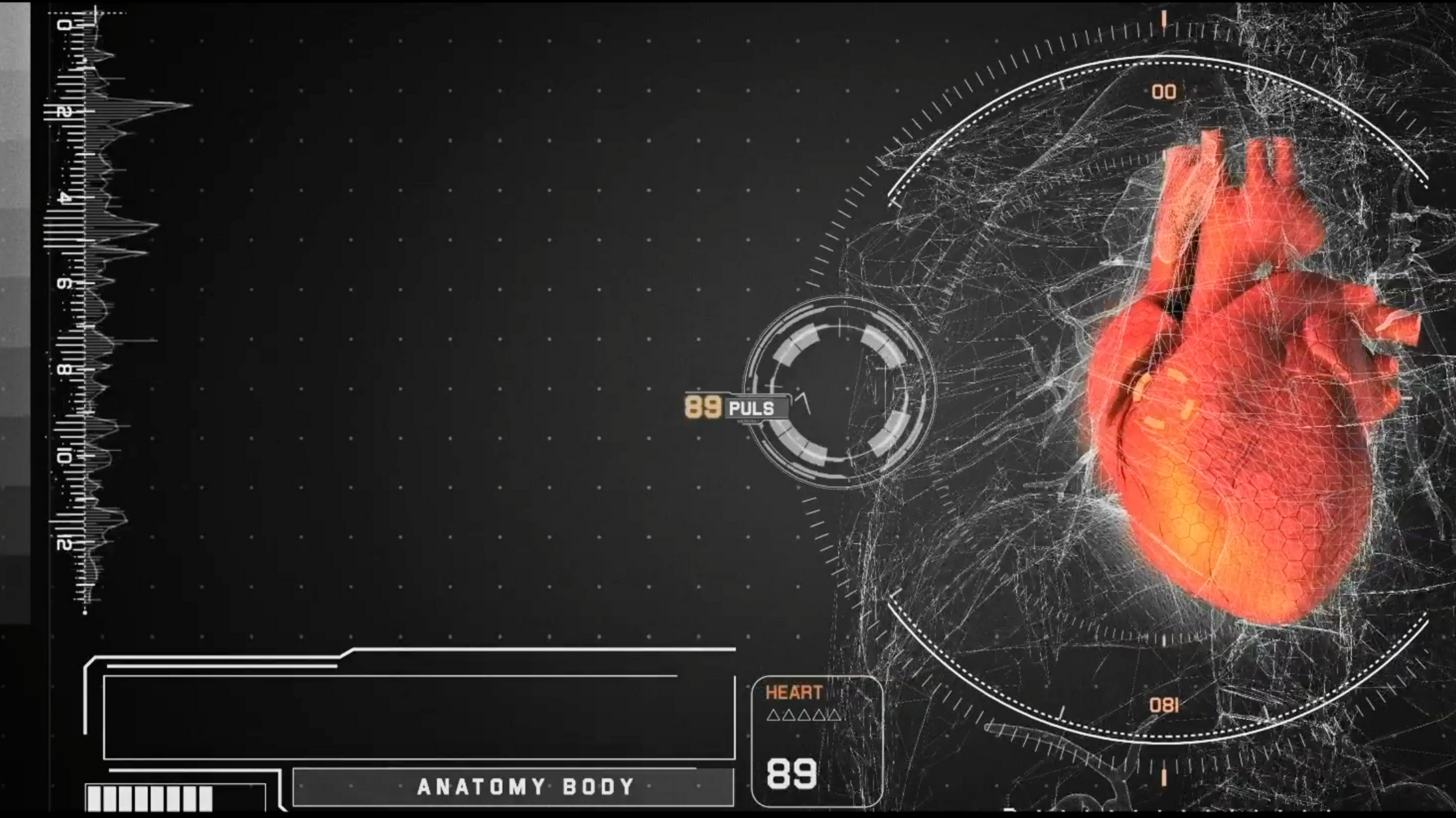
Way back in 1992, corpuls brought the 12-lead ECG to the pre-clinical market with their revolutionary and award winning corpuls 08/16 device. Now, nearly 30 years later, corpuls are doing it again with the ability to see 22 Leads, from the same 10 electrodes with their ECGmax.
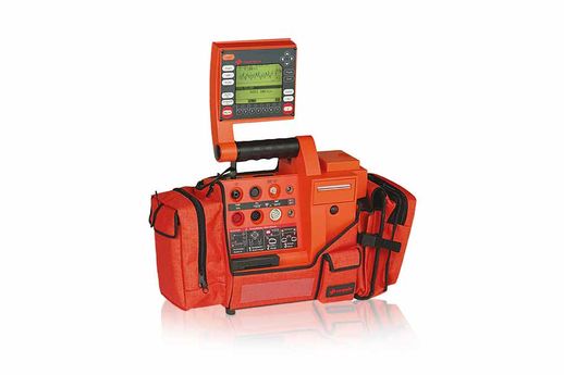 The 1992 corpuls 08/16 model
ECGmax
With the ECGmax you not only get the usual 12 leads, you get an additional 10 and thus a much more comprehensive picture of the heart muscle, which importantly includes its right side and the posterior wall. Monitoring of these aspects is recommended by the European Society of Cardiology guidelines wherever possible. Beneficially, to get this measure, no additional effort is required and no extra electrodes have to be attached or positioned by the user - all the computation is done server side.
ECGmax provides users with;
> Broadened diagnostics with 22 leads
> Posterior leads V7–V9
> Right cardiac leads V3r–V6r
> Orthogonal leads X, Y, Z and Vectorloops
> Only 10 electrodes, extremities and chest leads
Vectorloops
Vectorloops consider the electrical conduction in the heart as a rotating dipole. The course of the peak of this vector is spatially represented as a loop in a three-dimensional individual coordinate system. The loops correspond to the P wave, the QRS complex and the T wave.
With a healthy myocardium, the loops are regular and "fluid looking", as opposed to a myocardium with a dysfunction, where the conduction of the dipole is irregular and jagged.
> Electrical conduction as a rotating dipole
> Represented as a loop in the 3D coordinate system
> Loops correspond to the P wave, the QRS complex and the T wave
> Healthy loops are more regular and "flowing"
> Dysfunction shows as irregular and jagged
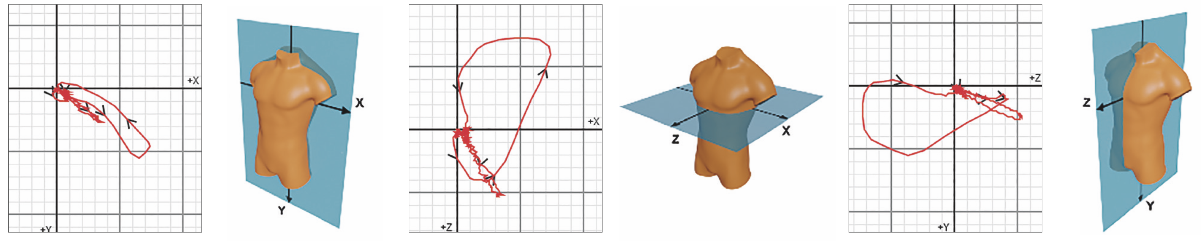
Visualization of the electrical activity of the three ortogonal leads (X,Y,Z) in the shape of vector loops
The Cardiac Electrical Biomarker
In addition, ECGmax can calculate the Cardiac Electrical Biomarker CEB® from the same electrodes. The three colour-coded areas of the CEB® – normal, conspicuous, abnormal – make the interpretation of myocardial ischemia particularly easy and the CEB® has comparable sensitivity and specificity to troponin.
> Simple interpretation using the traffic light concept
> Measured value is comparable to troponin
> Fast reaction by measuring the electric field
> High sensitivity and specificity
> No additional electrodes required
> Continuous value
> Non-invasive measurement
Watch the full explanation from the corpuls Innovation Summit (CIS) below 👇👌

Leave Comment