.jpg?width=342&height=509&name=OrtusAcademy(YELLOWGradient).jpg)


In this article, we'll explore how the ability to adjust ECG filters can help you maintain rhythm detection even when complex environments interfere with signals.
With the increase in diseases of the cardiovascular system, the electrocardiogram (ECG) is an essential tool for preclinical diagnostics in modern emergency medicine. It is recommended to continuously monitor patients with such diseases so to quickly detect life-threatening arrhythmias. [1]
In the preclinical setting, the recorded ECG may show artefacts due to various environmental factors. These can affect the interpretation of the ECG signal. [5] Artefacts can be caused by the patient’s muscle activity, transport movements or electrical coupling of the mains frequency, for example. To reduce the quantity of different artefacts in the ECG recording, corpuls devices offer the possibility to use different ECG filters.
The human ECG spectrum covers the frequency range 0.05 Hz - 100 Hz, with the main part of the QRS complex being in the range 0.5 Hz - 45 Hz. [9] Most of the time the ECG signal is overlaid by the mains frequency of 50 Hz/60 Hz. The mains frequency portion of an ECG can be reduced with a notch filter that can selectively filter individual frequencies. Low-pass filters are used to reduce high-frequency interference such as muscle tremors. These artefacts lie within the range from 5 Hz to 450 Hz. [10] High-pass filters in turn dim low-frequency interference signals that arise, for example, from breathing movements. These are between 0.05 Hz and 1 Hz. [11] This means that high and low frequency interference overlaps the frequency range of the ECG signal. Due to these technical reasons, filtering not only reduces interference, but also changes the ECG morphology. [3] The combination of low- and high-pass filters is known as a bandpass filter.
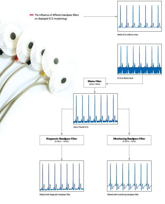
The principle of ECG filtering is shown in Figure 1. First, the mains frequency in the ECG is reduced with a notch filter. The notch filter must be set to the local mains frequency.
The intended use plays a decisive role in the selection of the bandpass filter. For diagnostic interpretation, the ECG signal must not be affected by the filtering. Therefore, it is recommended for diagnostic purposes to use a high-pass filter with 0.05 Hz and a low-pass filter with 150 Hz. [4] With this bandpass filter setting, the ECG is displayed with the maximum available frequency bandwidth. If the ECG is displayed with a reduced frequency bandwidth, the ECG morphology will be changed, for example, adjustment to the low-pass filter affects the amplitude of the R wave and adjusting the high-pass filter can result in shifts in the ST segment. [3] If the bandpass filter impairs the interpretation ability of the ECG signal, it will be shown on the ECG printout.
The aim of ECG monitoring is the detection of life-threatening arrhythmias such as ventricular fibrillation, ventricular tachycardia or asystole. [1] The detection of such rhythms can be impaired by various interferences during patient transport. [7] If rhythm detection is no longer possible, the bandpass filter with the smallest frequency bandwidth should be used. With this filter setting, the maximum possible number of interference frequencies is filtered from the ECG signal, and whilst the ECG is shown with less noise, it can only be used for rhythm analysis. [8]
Figure 1: How Filtering Influences the ECG Morphology
Corpuls
Download the corpuls ECG reference sheet here.
What to learn more about the ECG capabilities of the corpuls system? ECGmax allows clinicians to view 22 leads from the same 10 electrodes. Click here to learn more.
References
[1] Ibanez B, James S, Agewall S, et al. 2017 ESC Guidelines for the management of acute myocardial infarction in patients presenting with ST segment elevation: The Task Force for the management of acute myocardial infarction in patients presenting with ST segment elevation of the European Society of Cardiology (ESC). Eur Heart J. 2018;39(2):119-177. doi:10.1093/eurheartj/ehx393
[2] Morley AP. Prehospital monitoring of trauma patients: experience of a helicopter emergency medical service. Br J Anaesth. 1996;76(5): 726- 730. doi:10.1093/bja/76.5.726
[3] Satija U, Ramkumar B, Manikandan MS. A Review of Signal Processing Techniques for Electrocardiogram Signal Quality Assessment. IEEE Rev Biomed Eng. 2018;11:36-52. doi:10.1109/ RBME.2018.2810957
[4] Thakor NV. From Holter monitors to automatic defibrillators: developments in ambulatory arrhythmia monitoring. IEEE Trans Biomed Eng. 1984;31(12):770-778. doi:10.1109/ TBME.1984.325237
[5] Levkov C, Mihov G, Ivanov R, Daskalov I, Christov I, Dotsinsky I. Removal of power-line interference from the ECG: a review of the subtraction procedure. Biomed Eng Online. 2005;4:50. Published 2005 Aug 23. doi:10.1186/1475-925X-4-50
[6] T. M. Chieng, Y. W. Hau and Z. Omar, “The Study and Comparison between Various Digital Filters for ECG Denoising,” 2018 IEEE-EMBS Conference on Biomedical Engineering and Sciences (IECBES), Sarawak, Malaysia, 2018, pp. 226-232, doi: 10.1109/IECBES.2018.8626661
[7] U. Satija, B. Ramkumar and M. S. Manikandan, “A Review of Signal Processing Techniques for Electrocardiogram Signal Quality Assessment,” in IEEE Reviews in Biomedical Engineering, vol. 11, pp. 36-52, 2018, doi: 10.1109/RBME.2018.2810957.
[8] Buendía-Fuentes F, Arnau-Vives MA, Arnau-Vives A, et al. High-Bandpass Filters in Electrocardiography: Source of Error in the Interpretation of the ST Segment. ISRN Cardiol. 2012;2012:706217. doi:10.5402/2012/706217
[9] Kligfield P, Gettes LS, Bailey JJ, et al. Recommendations for the standardization and interpretation of the electrocardiogram: part I: the electrocardiogram and its technology a scientific statement from the American Heart Association Electrocardiography and Arrhythmias Committee, Council on Clinical Cardiology; the American College of Cardiology Foundation; and the Heart Rhythm Society endorsed by the International Society for Computerized Electrocardiology. J Am Coll Cardiol. 2007;49(10): 1109-1127. doi:10.1016/j.jacc.2007.01.024
[10] Yun JG, Jeung KW, Lee BK, et al. Performance of an automated external defibrillator in a moving ambulance vehicle. Resuscitation. 2010;81(4):457-462. doi:10.1016/j.resuscitation.2009.12.031
[11] N. V. Thakor and Y. -. Zhu, “Applications of adaptive filtering to ECG analysis: noise cancellation and arrhythmia detection,” in IEEE Transactions on Biomedical Engineering, vol. 38, no. 8, pp. 785-794, Aug. 1991, doi: 10.1109/10.83591.

Leave Comment